tree in bud opacities in lungs
Multiple causes for tree-in-bud TIB opacities have been reported. Cryptogenic organizing pneumonia 119.

Chest Imaging In The Diagnosis Of Occupational Lung Diseases Clinics In Chest Medicine
We here describe an unusual cause of TIB during the COVID-19 pandemic.

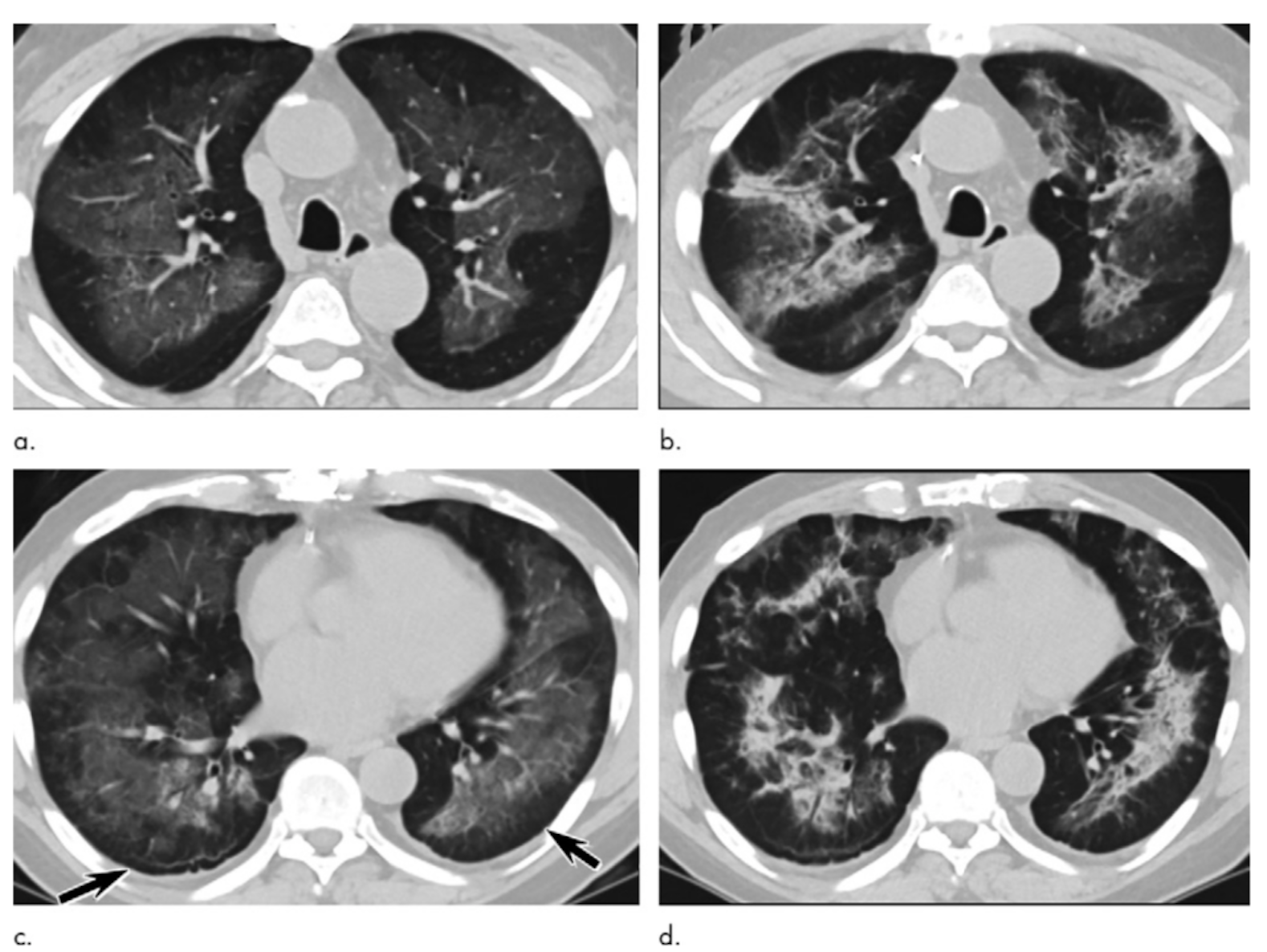
. The tree-in-bud pattern can be an early sign of disease Fig 10 15. Nodular opacities with tree-in-bud appearance can be associated with other changes in lung parenchyma-such as thickening of the bronchial walls consolidations andor areas of. Miller WT Jr Panosian JS.
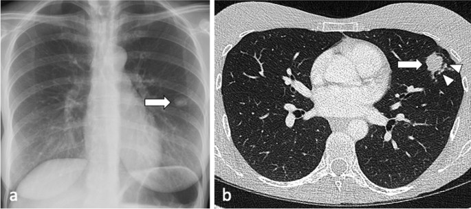
Computerized detection of tree-in-bud pattern. The tree-in-bud pattern is commonly seen at thin-section computed tomography CT of the lungs. A young male patient who had a history of fever cough and respiratory distress presented in the emergency department.
Usually somewhat nodular in appearance the tree-in-bud pattern is generally most pronounced in the lung periphery and associated with abnormalities of the larger airways. Although initially described in 1993 as a thin-section chest CT finding in active tuberculosis TIB opacities are by. Multiple causes for tree-in-bud TIB opacities have been reported.
The purpose of this study was to determine the relative frequency of causes of tib opacities and identify patterns of disease associated with tib opacities. The tree-in-bud sign is a nonspecific imaging finding that implies impaction within bronchioles the smallest airway passages in the lung. 11 TIB opacities represent a central imag- Background.
These small clustered branching and nodular opacities represent terminal airway mucous impaction with adjacent peribronchiolar inflammation. Cytomegalovirus pneumonia in a 51-year-old man with chronic myelogenous leukemia who underwent bone marrow transplantation. Tree-in-bud TIB opacities are a common imaging finding on thoracic CT scan.
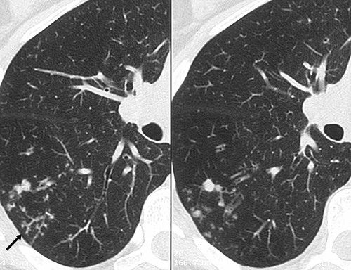
A Thin-section CT scan of the right lung shows centrilobular ground-glass opacities in addition to nodules and tree-in-bud opacities arrow. Not only are these patterns difficult to detect but micro-nodules and other normal and abnormal structures have strong shape and appearance similarities with existing structures in the lungs. As in this case renal cell carcinoma is one of the most common malignancies that may produce this vascular cause of tree-in-bud pattern.
In radiology the tree-in-bud sign is a finding on a CT scan that indicates some degree of airway obstruction. 1 2 3 4 Reported causes include infections aspiration and a variety of inflammatory conditions. The tree-in-bud-pattern of images on thin-section lung CT is defined by centrilobular branching structures that resemble a budding tree.
These small clustered branching and nodular opacities represent terminal airway mucous impaction with adjacent peribronchiolar inflammation. However to our knowledge the relative frequencies of the causes have not been evaluated. The tree-in-bud sign is a nonspecific imaging finding that implies impaction within bronchioles the smallest airway passages in the lung.
In radiology the tree-in-bud sign is a finding on a CT scan that indicates some degree of airway obstruction. Another important entity that can produce the tree-in-bud pattern is bronchioalveolar carcinoma BAC 1. TIB opacities represent a normally invisible branches of the bronchiole tree 1 mm in diameter that are severely impacted with mucous pus or fluid with resultant dilatation and budding of the terminal bronchioles 2 mm in diameter1 photo.
Subsequently question is what does tree in bud mean. It consists of small centrilobular nodules of soft-tissue attenuation connected to multiple branching linear structures of similar caliber that originate from a. Lung nodules are usually about 02.
3 found that the tree-in-bud pattern was seen in 256 of the CT scans in patients with bronchiectasis. TIB opacities are also associated with bronchiectasis and small airways obliteration resulting in mosaic air trapping. In radiology the tree-in-bud sign is a finding on a CT scan that indicates some degree of airway obstruction.
The tree-in-bud sign is a nonspecific imaging finding that implies impaction within bronchioles the smallest airway passages in the lung. The purpose of this study was to determine the relative frequency of causes of TIB opacities and identify patterns of disease associated with TIB opacities. This is the classic appearance of the tree in bud pattern seen on chest ct.
1 direct filling of the centrilobular arteries by tumor emboli and 2 fibrocellular intimal hyperplasia due to carcinomatous endarteritis. AJR Am J Roentgenol 1998171365370. What does tree-in-bud opacities mean.
In radiology the tree-in-bud sign is a finding on a CT scan that indicates some degree of airway obstruction. Rossi SE Franquet T Volpacchio M Gimenez A Aguilar G. This collage is presented to reveal tree in bud changes resulting from impaction in the smaller terminal bronchioles and respiratory units.
We investigated the pathological basis of the tree-in-bud lesion by reviewing the pathological specimens of bronchograms of normal lungs and contract radiographs of the post-mortem lungs manifesting active. In radiology the tree-in-bud sign is a finding on a CT scan that indicates some degree of airway obstruction. 1 5 6 7 8 9 10 11 12.
What does tree-in-bud opacities mean. Tree in Bud Sign Bronchopulmonary Aspergillosis ABPA CT scan through the chest shows medium sized bronchi bronchioles and small airways impacted with fluid. Tree-in-bud TIB opacities are a common imaging finding on thoracic CT scan.
However to our knowledge the relative frequencies of the causes have not been evaluated. Clin Chest Med 201536299312 ix. Tree-in-bud TIB appearance in computed tomography CT chest is most commonly a manifestation of infection.
Causes and imaging patterns of tree-in-bud opacities. Bronchiectasis which may be of any cause can produce the tree-in-bud pattern. Richards JC Lynch DA Chung JH.
Tib opacities are also associated with bronchiectasis and small airways obliteration resulting in mosaic air trapping. The tree-in-bud sign is a nonspecific imaging finding that implies impaction within bronchioles the smallest airway passages in the lung. There are many technical obstacles to detecting complex shape patterns such as tree-in-bud that are associated with pulmonary infections.
Is a 7mm lung nodule big. The tree-in-bud sign is a nonspecific imaging finding that implies impaction within bronchioles the smallest airway passages in the lung. Tree-in-bud refers to a pattern seen on thin-section chest CT in which centrilobular bronchial dilatation and filling by mucus pus or fluid resembles a budding tree.
Cystic and nodular lung disease. A tree-in-bud pattern of centrilobular nodules from metastatic disease occurs by two mechanisms.

Ct Scan Of Chest Revealing Scattered Tree In Bud Opacities In Both Download Scientific Diagram

Tree In Bud Pattern Pulmonary Tb Eurorad

Diffuse Pulmonary Calcifications A Case Series And Review Of Literature Jarjou I 2021 Respirology Case Reports Wiley Online Library

Tree In Bud Sign And Bronchiectasis Radiology Case Radiopaedia Org

P201 Mediastinal Hodgkin Lymphoma The Answer To Post 200 Swipe Left For Other Images Thanks For The Comm Pediatric Radiology Hodgkins Lymphoma Lymphoma

High Resolution Chest Ct Scan Axial Slice Shows Tree In Bud Pattern Of Download Scientific Diagram

Hrct Of The Lung Signs Of Infection Bilaterally Tree In Bud Patterns Download Scientific Diagram

Ground Glass Opacity Lung Nodules In The Era Of Lung Cancer Ct Screening Radiology Pathology And Clinical Management
Active Pulmonary Tuberculosis Something Old Something New Something Borrowed Something Blue Insights Into Imaging Full Text
Ct Scans Show Vaping Related Lung Injury Patterns

References In Causes And Imaging Patterns Of Tree In Bud Opacities Chest
Tree In Bud Sign An Overview Sciencedirect Topics

Infectious Bronchiolitis With Extensive Tree In Bud Pattern Radiology Case Radiopaedia Org Pattern Bud Case
View Of Tree In Bud The Southwest Respiratory And Critical Care Chronicles
Tree In Bud Sign Lungs

Plain Film Appearance Of Bronchiectasis In Kartagener S Syndrome A Pa Chest Radiograph Shows Situs Inversus Increased In 2022 Situs Inversus Bronchial Radiographer

Signs Of Bronchiectasis Tram Tracks Thick Rings Signet Ring Sign And Finger In Glove Sign This Is Cystic Fibros Medical Ultrasound Radiology Imaging Pet Ct

Pin Em Radiologia

Pin On Ct Scans